Functional Magnetic Resonance Imaging (MRI) changes in Type 1 Diabetes with and without Renal Hyperfiltration (#221)
Introduction
We aimed to determine if functional MRI is able to detect change in renal function in patients with type 1 Diabetes (T1DM).
We hypothesized that compared to controls, patients with T1DM have:
(i)a decrease in intra-renal oxygenation as measured by blood oxygen level dependent (BOLD) imaging, represented by an increased medullary to cortical R2* (MCR), and
(ii)changes in diffusion tensor imaging (DTI) metrics, fractional anisotropy (FA) and apparent diffusion coefficient (ADC), representing renal parenchymal water motion.
We further hypothesized that changes in T1DM patients are more pronounced in patients with compared to those without hyperfiltration.
Methods
32 patients with T1DM and 10 healthy controls underwent 3-Tesla MRI in a prospective study.Hyperfiltration was defined as a measured Glomerular Filtration Rate (mGFR)≥120mls/ min/1.73m2, and Stage1+ Diabetic Kidney Disease(DKD)as mGFR<120mls/ min/1.73m2 determined by 99mTc--diethylene-triamine-pentaacetate(DTPA).Based on intra and inter-reader concordances from two blinded readers (R1 and R2), we performed analyses on BOLD data for both readers and DTI data for R1 alone. MCR, FA and ADC values between: (i)T1DM versus Controls (ii)T1DM with hyperfiltration versus those without were analysed using Mann-Whitney tests.
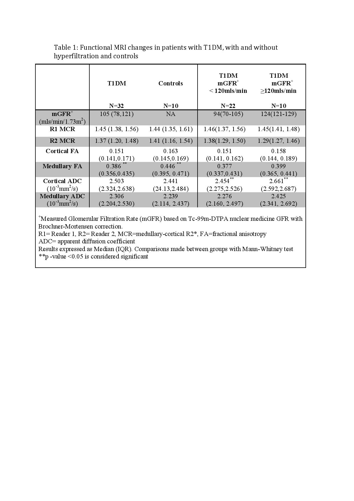
Results (Table 1)
There were no significant differences between T1DM and controls,except for medullary FA(p=0.03).Lower medullary FA in the stage1+DKD group compared to hyperfiltration did not reach statistical significance(p=0.4).T1DM with hyperfiltration had a significantly higher cADC compared to those with stage1+DKD(p=0.0007).No differences were found between hyperfiltration and stage1+DKD for BOLD-derived parameters.
Conclusion:
There were no differences in functional MRI parameters between T1DM and controls apart from a lower mFA in T1DM.This finding, together with the lower cADC in the stage1+DKD group may potentially reflect disruption of medullary tubular architecture in progressive DKD. These findings should be interpreted with caution given the small patient population.Further studies in a larger cohort may demonstrate differences between different degrees of DKD using functional MRI.